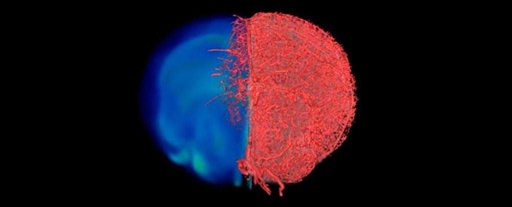
New imaging technique shows blood vessels
Blood vessels are pretty important when it comes to the healthy functioning of the body, and researchers and health professionals need to know as much as possible about where these tiny transport channels are going.
A newly developed 3D visualization technique should help. It's called VascuViz, and it uses a quick-setting polymer mixture that fills up blood vessels and makes them visible to a wide variety of scanning technologies as they move around tissues and organs.
Lab mouse tests so far indicate it works at different scales – from the largest arteries to the smallest capillaries – and can be used to show details that would otherwise get missed by conventional techniques, improving our understanding of how tissues work.
Measurements taken by VascuViz can then be entered into computer simulations of blood flow, including cancer models, to figure out how blood is flowing – something that's essential in understanding how diseases work and might be progressing.
What makes the new technique so useful is that it offers an all-in-one approach, providing results and a level of detail that would normally take several scans and several different methods to achieve.
Existing imaging methods like magnetic resonance imaging (MRI),
computed tomography (CT) and microscopy all have their roles in studying blood vessels in the lab, but they don't work all that well together and must be run separately.
The VascuViz technique uses a combination of imaging agents: BriteVu (used in CT scans) and Galbumin-Rhodamine (used in MRI scans). What's then produced is a wonderfully detailed, three-dimensional model of blood vessel position.
Cancer tumours, leg muscles, the brain, kidney tissues and circulatory system could all be imaged by VascuViz.
 English
 Arabic


